Learn More About Corneal Conditions
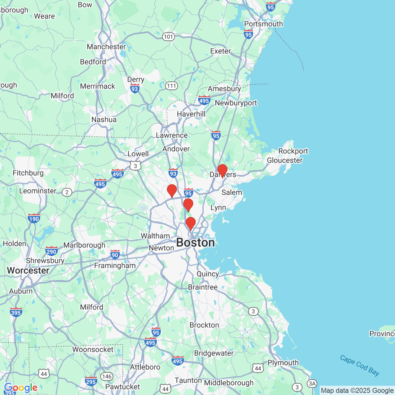
The dome-shaped, transparent protective layer covering the front of the eye, the cornea, plays an important role in eye health. In addition to providing a comprehensive range of services designed to improve and maintain patients' vision, Drs. Nilesh M. Sheth and Robert Kupsc of the Sheth-Horsley Eye Center provide diagnosis and treatment for conditions of the cornea for Boston area residents and surrounding communities.
To learn more about available treatments and procedures, or schedule an examination with one of our dedicated surgeons, reach out to our office today using our simple online contact form.
Corneal Transplant
Corneal transplants, either full thickness or back layer (also known as penetrating keratoplasty or PK), use an organ donor's healthy cornea to replace diseased, damaged or scarred corneal tissue. The two procedures differ in the amount of tissue that the graft replaces. Since an unhealthy cornea may result in distorted or blurred vision, a corneal transplant can effectively restore normal, functional vision.
Fuchs' Dystrophy
Fuchs' dystrophy occurs when endothelial cells deteriorate and the endothelium (the innermost layer of the cornea) swells and distorts vision. Pain and visual impairment become more pronounced as the epithelium takes on more water and the cornea's natural curvature is altered. Treatment options include soft contacts, prescription eye drops or ointments, and eventually, corneal transplant. The condition is typically an age-related disease afflicting patients in their 50's and over.
Post LASIK Ectasia
Ectasia occurs when the natural curvature of the cornea becomes progressively steeper. Since LASIK permanently thins and reshapes the covering of the eye, post-LASIK ecstasia has been observed as a complication of the refractive surgery. As the cornea becomes more cone-shaped and bulges outwards, vision deteriorates and may eventually necessitate a corneal implant.
Keratoconus
Similarly to ecstasia, keratoconus involves the steepening of the cornea's natural shape. As the cornea progressively bulges outwards, visual distortion occurs in the form of blurriness, sensitivity to bright lights and difficulty seeing at night. Typically diagnosed in patients 10-25 years old, treatment includes glasses, rigid gas permeable contacts and corneal transplant.
Descemet’s Stripping Automated Endothelial Keratoplasty (DSAEK)
Implemented as an alternative to penetrating keratoplasty (PK) in which the entire thickness of the cornea is replaced with donor tissue, DSAEK, also known as partial thickness corneal transplant, targets the endothelial layer of the cornea. Since only the inner layer of the cornea is replaced, benefits include improved healing time and decreased risk of complications surrounding the transplant tissue.
Pterygium
A pterygium is made up of a fleshy, raised growth that grows on the white of the eye. Also known as a scleral conjunctiva, a pterygium is generally harmless but sufferers may wish to have it removed for cosmetic reasons. Advanced pterygia may cause astigmatism and can be treated through steroid eye drops or surgery, if needed.
Corneal Scar
Though the cornea displays remarkable healing properties and resiliency to injury, corneal scars may be caused by a number of factors including trauma, infection or disease. If vision is severely affected, a corneal transplant may be performed to restore clarity of vision.
Cogan’s Dystrophy (Map-Dot-Fingerprint Dystrophy)
Cogan's Dystrophy is a disorder of the cornea resulting in progressive dystrophy, or thinning of the lens. Also known as map-dot-fingerprint dystrophy, Cogan's Dystrophy causes damage to the epithelial or outermost layer of the cornea, in which cells tear away from surrounding tissue and cause red eye, blurred vision and pain. Treatment options range from prescription eye drops to corneal transplant, depending on the severity of the condition.


